‘Google Maps’ for the body: program uses zoom technology to image entire organs down to cellular level
Technology was top-secret for some time, has been formerly presented to research society
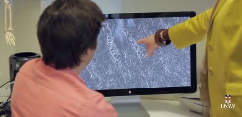
A collaborative team of researchers in Australia has formerly presented a top-secret biomedical technology they’ve been working on for some time.The best way to describe it is “Google Maps” for the human body — using Google algorithms, the researchers can zoom in and out from the scale of a whole organ down to the cellular level “just as you would with Google Maps”, reducing to “a matter of weeks analyses that once took 25 years to completed.”
What’s particularly fascinating about this story is that the imaging technology was developed by German optical and industrial measurement manufacturer Zeiss for the purpose of scanning silicon wafers for defects.
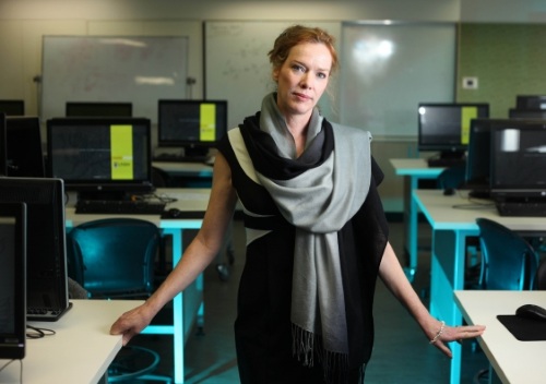
University of New South Wales professor Melissa Knothe Tate, the Paul Trainor Chair of Biomedical Engineering and the person who led this project, re-thought the technology’s purpose as being appropriate for exploring biological issues; specifically, osteoporosis and osteoarthritis.
Using Zeiss’s cutting-edge microtome and MRI technologies, coupled with Google’s industry-leading digital map programming, Tate’s team can examine with incredible detail how movement and weight bearing affects the movement of molecules within joints. They’re also able to explore the relationship between blood, bone, lymphatics, and muscle.
"For the first time we have the ability to go from the whole body down to how the cells are getting their nutrition and how this is all connected," said Ms. Tate. "This could open the door to as yet unknown new therapies and preventions."
The one thing that’s been holding the industry back is the development of a microscopy technology that allows seamless imaging of organs and tissues across length scales, as well as the ability to go through and study the enormous amount of data that comes with an analysis.
The application of semiconductor imaging technology on humans marks the first time such an instance has occurred. It is believed that by understanding the molecular signaling and traffic between tissues, the range of treatments will not only be expanded, but more effective too.
"These are terabyte-sized data sets so the Google maps algorithms are helping us take this tremendous amount of information and use it effectively. They're the traffic controllers, if you like," Tate explained. "Advanced research instrumentation provides a technological platform to answer the hardest, unanswered questions in science, opening up avenues for fundamental discoveries, the implications of which may be currently unfathomable yet which will ultimately pave the way to engineer better human health and quality of life as we age."
Ms. Tate has since established partnerships with the US-based Cleveland Clinic as well as Brown and Stanford Universities. Zeiss and Google have also gotten involved in the project, by helping to crunch terabytes of data gathered from human hip studies.
To see the technology in action, and to hear Ms. Tate explain it a bit further, check out the clip below:
Via the University of New South Wales
Learn more about Electronic Products Magazine
Copyright © by Hearst Business Communications, Inc. All Rights Reserved.
http://www.electronicproducts.com/Biotech/Research/Google_Maps_for_the_body_program_uses_zoom_technology_to_image_entire_organs_down_to_cellular_level.aspx