Camera-loaded catheter streams live from inside arteries, removes plaque at same time

The Pantheris system can not only spot blockages in blood vessels, but it can help doctors shave them away as well
The impact of ever-miniaturizing electronics can be felt right across the spectrum of technological advancement, but as we are beginning to see, one place where it can have a truly profound impact is in the human body. The latest example of this is a tiny camera no bigger than a grain of salt, which can be fixed to the end of a catheter and fed into arteries to provide surgeons tasked with removing plaque a live view from within.
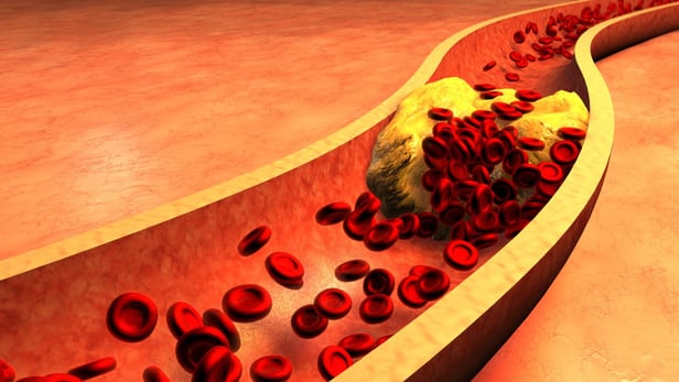
The buildup of plaque inside blood vessels can lead to all sorts of health problems, the most extreme of which are life-threatening events like heart attacks and strokes. Doctors try to intervene before things get this bad by cutting plaque from the artery to improve blood flow, after first imaging the clogged vessel using techniques like x-ray to size up the job at hand.
But the trouble with this approach is that it only provides doctors with a cross-section view of the vessel. You could think of it like trying to clear a blockage in a pipe while only being able to see that pipe from the side. But lately, scientists are coming up with tiny technologies that allow us to venture inside for a look around.
Earlier this year we reported on a new 3D-printing technique that allowed scientists to produce a complex optical lens system for medical imaging that was around the size of a grain of salt and could be loaded into a syringe. And back in 2014, scientists developed a tiny catheter-based sensor that could provide a real-time view from within blood vessels to offer doctors a better vantage point.
And now, this approach is finding its way into the clinic as part of real-world procedures. The Pantheris Lumivascular atherectomy system device one ups the aforementioned solutions because it not only provides live images from inside, but doctors can use it to clear out the vessels at the same time.
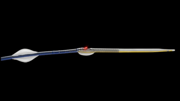
After being fed through an incision in the groin, Pantheris uses optical coherence tomography (OCT), a medical-imaging technique that relies on light waves to gather high-res images from within biological material, to provide doctors with a live view from inside the artery. Simultaneously, doctors can engage a cutting mechanism that is also fixed to its tip to shave off plaque as needed, using the real-time images for guidance.
After it received FDA approval back in March, doctors at UC San Diego Health have used Pantheris on ten patients. Most recently, it was deployed to clear an artery of a patient with severe scar tissue and buildup of plaque at a previously treated site. With the blockage located in his right leg, prior to treatment the patient had trouble walking as a result of limited blood flow to his calf muscle. But following the successful procedure, the patient is now able to walk several miles at a time.
"He was a good candidate for the new image-guided catheter approach," says Mitul Patel, MD, cardiologist at UC San Diego Health. "The device allowed for excellent visualization inside his leg artery as we removed only the diseased tissue."
You can watch the device in action in this video.
Source: UC San Diego Health, Avinger